Case of the Week #4 – Closed Pyometra
Pet: Sophie
Age: 7 yrs, 4 mos.
Species: Canine
Breed: Vizsla mix
Color: Red
Sex: Female, intact
Wt. on presentation: 56 lbs
Sophie presented to us for the first time in November 2009, with a history of acute onset of extreme lethargy, anorexia, vomiting, and small bouts of diarrhea. The problem first appeared two days prior, but she deteriorated rapidly throughout the previous day, and on the morning of the presentation was very weak.
The history for Sophie prior to her current condition was relatively normal. She had been eating and drinking completely normally, with no vomiting or diarrhea, and was very active and playful. She was not current on vaccines and was not on any flea or heartworm control. She had been in heat about 6 weeks earlier, during which she was definitely not bred.
Her physical exam (PE) was interesting. Her temperature was 98.6. The normal temperature in a dog is 101 to 102.5, so hers was definitely sub-normal. Her pulses were strong to bounding, and her heart rate was 156. For a dog her size, a normal heart rate is about 100 – 120 bpm.
Her abdomen was full to bloated, to the point where I couldn’t identify any of the usual internal structures, such as bladder, liver, or spleen. And even though her abdomen was “full” (the owners thought she looked “fat”), careful examination of her spinal muscles showed she was actually muscle wasting and losing weight, not gaining it.
Her heart auscultated normally, with no murmurs or arrhythmias. However, as with the pulses, her heart had a bounding character to it. And as stated above, she had a higher than normal heart rate. Her lungs were clear in all quadrants.
Her peripheral lymph nodes were all normal, and her eyes, ears, nose, and throat were all normal with the exception of her eyes being possibly sunken in mildly. Her gums were normal pink, and when pressed they turned white, then returned to pink in two seconds, which is normal.
Given the history, the number one differential diagnosis by far was a closed pyometra. In short, however, pyometras are severe infections in the uterus. They tend to occur about 2 weeks to 2 months after a bitch has her heat cycle. And if the cervix is closed, the uterus fills rapidly with pus, making the dog extremely ill very fast. In this case, the decreased body temperature, increased heart rate, and bounding heartbeat all pointed to the probability of septic shock.
Lab work was run and abdominal films were shot. The total white blood cell count was actually at the high end of normal. Given the overwhelming infection involved in pyometra, we always look for a high white cell count to confirm. However, in some cases – this one included – the infection and depression come on so suddenly that the body doesn’t get a chance to react before we test the blood. So in this case, the finding of a high-normal white cell count did not in any way rule out pyometra. The rest of the lab work showed that the pet was moderate to severely dehydrated, but otherwise, her cell lines and organ functions were normal.
Abdominal x-rays confirmed a large, tortuous tubular structure in the entire ventral (lower) and caudal (rear part) abdomen. This was the uterus, which was completely filled with pus. In a normal bitch the uterus is not visible on films at any part of the hormone cycle. The only time it’s visible is either when the bitch is pregnant, or during a pyometra. This was clearly a closed pyometra.
Closed pyos present a severe treatment challenge. As stated, this pet was very, very sick and headed straight into septic shock. That’s hardly an ideal surgical candidate. However, pyometra dogs never, ever get better with medicine alone. They always need surgery, and the sooner the better. So the goal, in this case, was to stabilize Sophie as much and as quickly as possible, and then to take her to surgery.
An IV catheter was placed, and a liter of fluid was administered immediately. IV antibiotics were given, along with anti-inflammatory and pain control injections. Two hours later she was sedated, prepped, and taken to surgery.
Here are some pictures from the surgery:
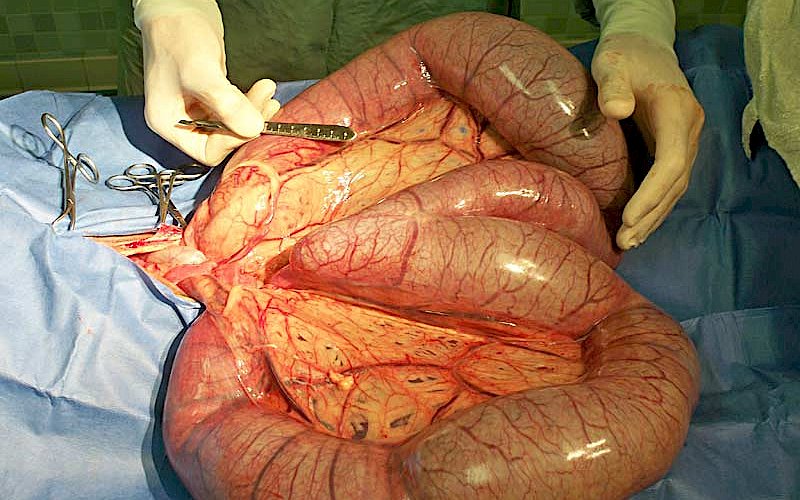
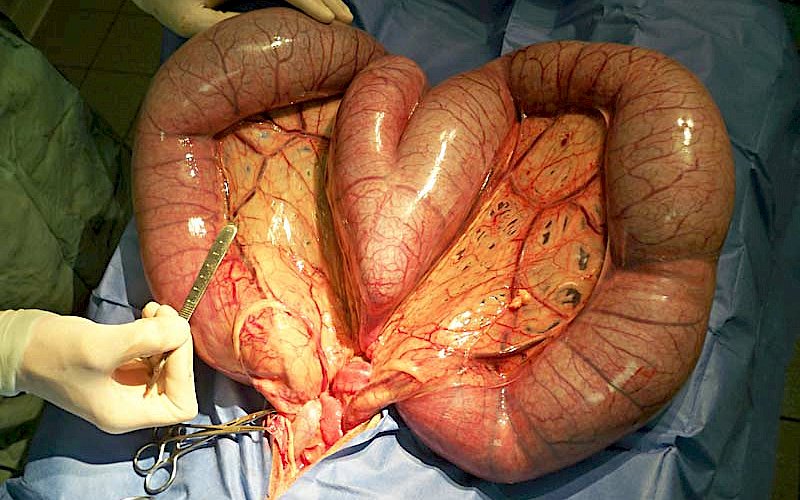
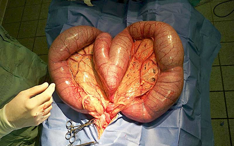

The uterus in dogs is not shaped like it is in women. In women, it’s shaped like a funnel or a triangle, where a single fetus implants and grows to maturity for birth. In dogs, the uterus is shaped like a wishbone. It has a small body adjacent to the cervix, which then bifurcates into two horns. These left and right horns are where the puppies line up during gestation and continue to grow and mature for birth.
When a dog Sophie’s size is not pregnant each of these two horns should be about the size of a normal #2 pencil. When pregnant, they’ll stretch and grow to be about 3 inches in diameter. In the adjacent pictures, you can see the horns after I have them exteriorized. Each one of the horns is bigger than my arm; these are completely full and stretched to the max by pus. We were very, very fortunate to get this pyometra out of the pet’s abdomen without it rupturing. Once we had it out it weighed over 11 lbs.
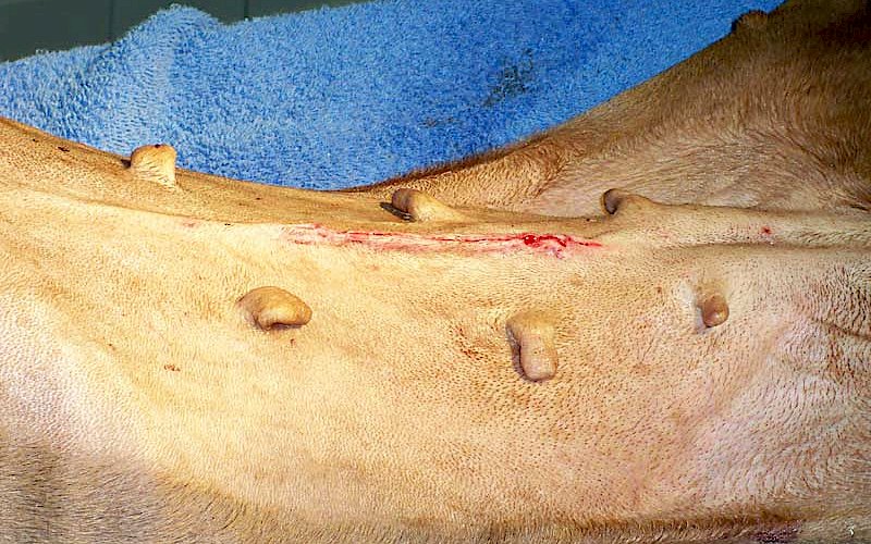
The uterus was removed, the abdomen flushed and closure was routine. Sophie was up and walking within a few hours, and was discharged from the hospital the next day. She made a full recovery.
Pyometras are extremely common, and when the cervix is closed, as in Sophie’s case, they represent an extreme emergency. Sophie was very fortunate. Other dogs are not so lucky. Pyometra is the number one reason why we recommend spaying all bitches not intended to be used for breeding.

Cookies on this website are used to both support the function and performance of the site, and also for marketing purposes, including personalizing content and tailoring advertising to your interests. To manage marketing cookies on this website, please select the button that indicates your preferences. More information can be found in our privacy policy here.