Case of the Week #3 – Tumor Removal
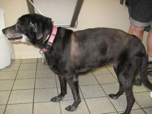
Pet: Angel
Age: 13 yrs, 7 mos.
Species: Canine
Breed: Labrador Retriever Mix
Color: Black
Sex: Female, spayed
Angel presented to Acupet Veterinary Care in May 2010 for evaluation of a lump on the pet’s left side. The mass had been there approximately 2 to 3 months, was 3 to 4 inches around, and was continuing to grow at the time of evaluation. The mass did not bother the dog; she did not appear painful, nor was there any scratching or chewing at the mass.
Upon physical exam the pet was determined to be in good overall condition. The abdomen palpated normally, heart and lungs were clear, peripheral lymph nodes were normal, and eyes/ears/nose/throat/mucous membranes were all normal for an 11-year-old dog.
The examined mass was approximately 4 inches round and 1 inch thick, located in the subcutaneous space between the pet’s skin and the underlying muscle wall. The mass was smooth and freely movable. One differential for the mass was a lipoma, which is a benign mass of fatty tissue origin. Lipomas are always benign, and never a problem. However this mass felt mildly firmer than a lipoma, so to determine whether we should be concerned we performed a fine needle aspirate (FNA).
The Fine Needle Aspirate
An FNA is a quick and painless procedure wherein we obtain a few cells from the area in question and examine them under the microscope. In the case of a lipoma, we should see few, if any cells, and instead see mostly just little drops of fat or oil on the slide. However, the FNA in Angel’s case revealed numerous abnormal red blood cells, which ruled out lipoma.
It was recommended at that time that we remove the mass surgically and send it in to the lab for analysis. The owner opted not to have it removed as it was slow growing and didn’t seem to be causing the pet any harm. He was sent home with no medications, and with instructions to inform us and have it removed should it continue to grow.
Over the next two years the mass did indeed continue to grow. At multiple different visits for other problems, it was reiterated to the owner that the mass was definitely a problem and needed to be removed. The owner continued to resist, and as the mass continued to grow the owner resigned himself that it was now at a point beyond the possibility of surgical intervention.
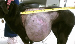
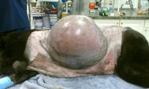
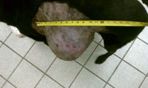
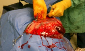
The pet was re-presented for evaluation of the mass in December 2012, 2.5 years after the initial consult on the mass. The mass was now approximately the size of a soccer ball protruding from the pet’s side. (See accompanying pictures below.) The owner was distraught, as he feared the mass was beyond surgical resection, and he now noticed that the mass was uncomfortable to the dog. After much discussion, we convinced him to allow us to try to remove it.
Tumor Removal Surgery
Angel returned 4 days later for surgery. Pre-anesthetic lab work was performed in-house and returned normal. Radiographs (x-rays) of the chest were taken to make sure the mass hadn’t spread to the lungs and were also normal. An IV catheter was placed and fluids started. The dog was sedated, pain control and antibiotic injections were given, and the site was prepped for surgery.
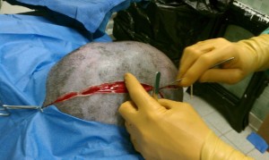
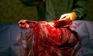
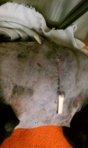
The surgery went extremely smoothly. (See accompanying pictures for pre-, intra-, and post-surgical pictures.) The mass was large, lobular, and extremely bloody. (Post-op the mass weighed in at 12.7 lbs.) However, it was well encapsulated and did not invade the underlying musculature. Along with the fact that the mass had been there two years and did not appear to spread, this was a good prognostic indicator that the mass was benign. The mass was removed along with a large wedge of the overlying skin, a sub-Q drain was placed to help prevent seroma or hematoma formation, and the area closed. Finally, a full body bandage was placed. Angel recovered from surgery without incident.
The mass was sent to the lab and did indeed come back as benign, with a guarded to a good prognosis. The drain was left in place for 5 days, then removed. The site was kept bandaged for 10 days, with bandage changes every 2 to 3 days. Sutures were removed 14 days post-op.
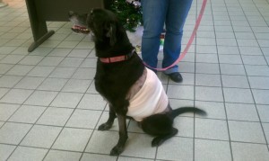
Angel has done extremely well since surgery. Her 3-month check-up (see pictures) shows no recurrence of the mass, and she is now more playful and energetic than she has been in years, according to the owner.
Angel was relatively lucky in that her mass never did spread or invade the body. Other dogs are not so lucky. Masses that are at first benign can, and often do, become much more of a problem the longer they’re left on. Early removal is always the best chance we have to make sure that tumors don’t become a non-manageable problem.
Angel’s photo gallery.

Cookies on this website are used to both support the function and performance of the site, and also for marketing purposes, including personalizing content and tailoring advertising to your interests. To manage marketing cookies on this website, please select the button that indicates your preferences. More information can be found in our privacy policy here.
We've upgraded our online store!
Ordering your pet's favorite food and medicine is now easier than ever.
Order Food & Meds
Quick & Easy Registration

Please use the phone number and email you currently use for hospital communications to link your account!
Linked Pet Records & Rx

Your pet's prescriptions and records will be waiting for you!
Pawsome
Savings!

AutoShip discounts, promotions on your favorite products and more!